

Cone beam computed tomography for complex developmental disorders
Imaging diagnostics are carried out using low-radiation digital X-rays or three-dimensional cone beam computed tomography (CBCT). In the case of complex developmental disorders of the teeth and jaws, diagnostics with a cone beam scanner provide a reliable basis for planning.
Even at the planning stage we obtain precise information on the bone situation and on the neighbouring anatomical structures. Cone beam computed tomography allows a spatial representation that is free of distortion and metrically accurate.
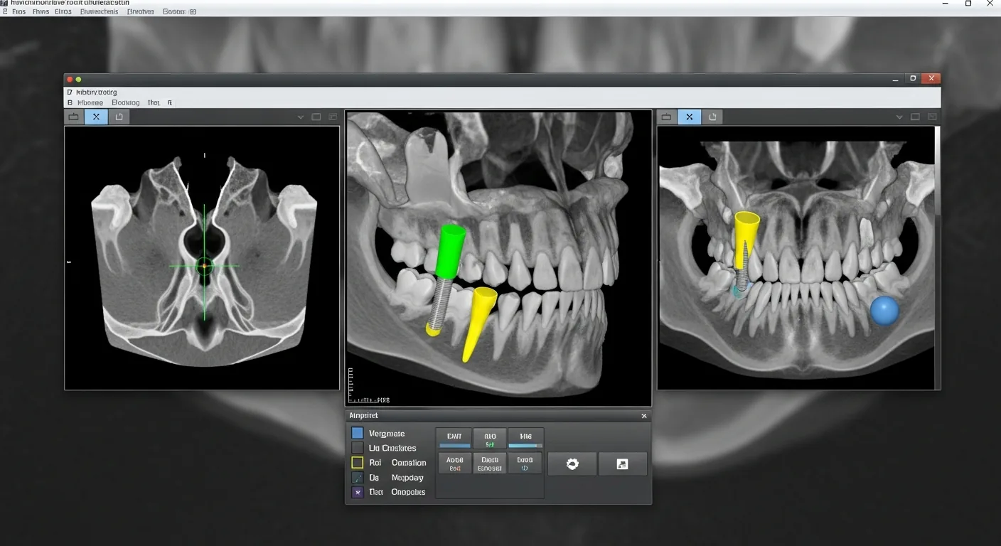
A detailed view of the structures of the jaw
The high-resolution CBCT images offer a detailed, three-dimensional view of the structures of the jaw and support both the treatment planning and the monitoring of the treatment.
We use three-dimensional diagnostics specifically where a two-dimensional image does not allow a sufficiently clear statement – that is, only when the diagnostic benefit justifies the effort.
For our patients, digital diagnostics mean treatment that is as gentle as possible and aesthetically demanding results.
Specialist orthodontic practice in Offenbach
Three-dimensional diagnostics in orthodontics
The use of three-dimensional diagnostics has considerably widened the examination options available in orthodontics and dentistry. The causes of unclear complaints can be located and analysed more effectively.
Low-radiation diagnostic procedures are the basis for a precise treatment plan.
We use 3D diagnostics in these areas:
- for complex malformations and misaligned teeth
- for anomalies of the teeth and tooth roots or disturbances of tooth eruption
- for retained and displaced teeth
- for pathological bone structures, cysts for example
The key facts at a glance
- Imaging diagnostics are carried out using low-radiation digital X-rays or cone beam computed tomography (CBCT).
- CBCT shows the jaw, tooth roots and neighbouring structures three-dimensionally, free of distortion and metrically precise.
- It is used for complex malformations, tooth and root anomalies, eruption disorders, displaced teeth and cysts.
- Three-dimensional images are taken selectively, only when a two-dimensional image does not permit a sufficient assessment.